
Howard Lecture: how human desire to render the invisible visible transformed the prostate cancer diagnosis pathway
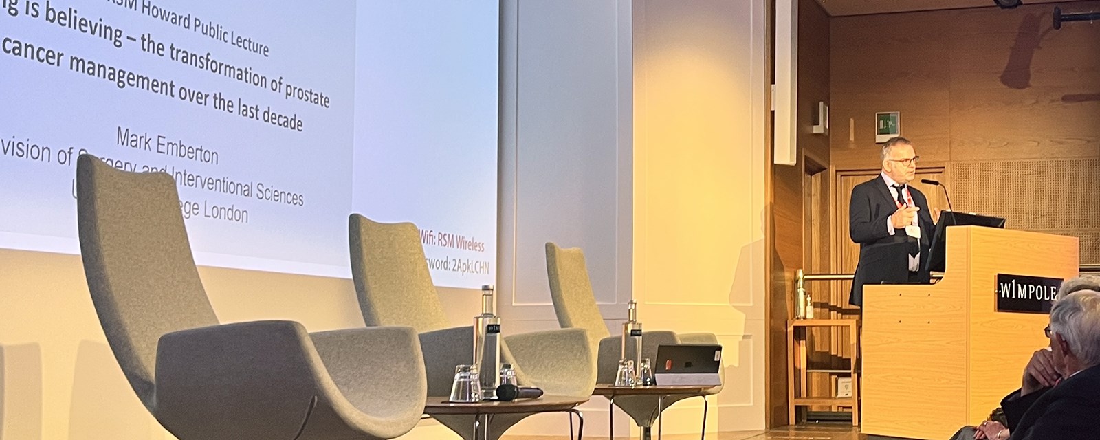
The Howard Foundation 40th anniversary lecture was given by Professor Mark Emberton, Professor of Intervention Oncology and Dean, Faculty of Medical Science at University College London, at the Royal Society of Medicine. The event was made free for all thanks to generous support from the Howard Foundation.
Prostate cancer is the most common cancer in men, with 52,000 new cases per year according to statistics from Cancer Research UK.
A live audience at the Royal Society of Medicine on 28 November heard how, until recently, the diagnosis pathway often led to harms. These included over and under treatment of patients, and high incidences of long-lasting side effects such as incontinence and impotence for those who were treated.
In 2019, NICE approved multi-parametric MRI scanning as a first-line investigation for prostate cancer diagnosis, ahead of biopsy. The change in guidance came about following two clinical trials led by UCL in partnership with University College London Hospitals (UCLH), with Professor Emberton at the helm.
Professor Emberton last week told a live audience at the 40th anniversary Howard Lecture the story of how the trials triggered the transformation of the diagnosis pathway.
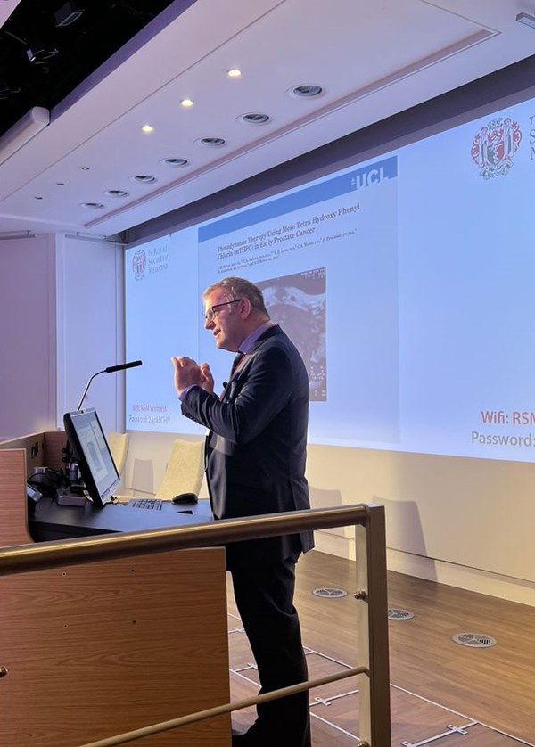
Professor Mark Emberton, Professor of Intervention Oncology and Dean, Faculty of Medical Science at University College London, delivering the Howard Foundation 40th anniversary lecture
But, as Professor Emberton said, the story is not about him alone. “This is a lot of people working together to solve problems and it’s been a privilege to have been part of it”.
The story begins on 14 May 1998 when, “huddled” around a “pretty blurry” early MRI image at UCLH, “the first moment of kind of wonder, and a sense of what might just be possible” occurred.
Professor Emberton explained: “We thought we’d do a sequence that they were doing in brain that had never really been done before in the prostate, and this was injecting contrast. We’d just treated this prostate with a new experimental technique, and we wanted to see whether we were making any difference to the tissue, to the prostate.
This was the first time in history that I think anybody had ever seen changes within the prostate that had resulted from attempt to treat the prostate.
“This was the first time in history that I think anybody had ever seen changes within the prostate that had resulted from attempt to treat the prostate. And we could only do that because MRI was just about ready and up for the task. And that was when we started thinking about the possibilities and the future.”
Prior to the introduction of MRI scanning, he said, the only way to ascertain what was going on in the prostate was through the rectum – with the finger being the “principal diagnostic tool” allowing doctors “to really feel what the prostate contained, or to ascertain its nature”. And, even then, “you could only access a really relatively small amount of the prostate – and we were reliant on that approach for a long, long time.”
Treatment options were limited to three approaches: no treatment; removal; or radiotherapy – all at the extremes of care. This led to many patients getting the wrong treatment or being undertreated, he said, as well as some patients being incorrectly given the all clear.
While every other cancer could be identified by a CT scan or an X-ray, those methods weren’t available for prostate cancer. Instead, if prostate cancer was suspected, to try to find it, urologists would “throw needles into the prostate in a random manner… without knowledge of the target that you were going after.” It was impossible to know the extent of the errors that were made with this “relatively unreliable” approach, Professor Emberton explained.
Identifying the potential for imaging to “render the invisible visible”, Professor Emberton’s unit set about to address the “unmet needs”, or “problems buried in the headlines” of the diagnosis and treatment methods of the time: how to ‘see’ the cancer; how to treat the cancer without the side-effects of incontinence and sexual dysfunction; and how to define the attributes of the cancer that benefits from treatment – allowing treatment to be tailored to the individual.
The world changed as a result.
The two clinical trials (PROMIS and PRECISION) led by UCL in partnership with University College London Hospitals (UCLH) and overseen by Professor Emberton, demonstrated that exposing patients to MRI prior to biopsy would result in health gains, including fewer missed cancers, better pathology and less harm. The gains also included “the clincher”: less cost, which prompted NICE to adopt the new approach, and “the world changed as a result”. The studies showed a quarter of a million men in Europe, if not more, would avoid unnecessary biopsy.
Professor Emberton cited collaboration across disciplines as one of the reasons policy change in the UK was able to be brought about more quickly than in other places. He said: “Because we all work together in teams and in groups there was no problem at all in sending a patient for an MRI - in fact, we would discuss it in groups and learn from each other in groups.”
The development of imaging enabled treatment to move away from the extreme of ‘whole gland’ treatment to the more tailored ‘focal therapy’ approach, saving tissue. The UCL research showed that “if we save a bit of tissue, men are 100% continent and men keep their erections, 95% [of the time].”
You can character the disease with amazing precision.
Professor Emberton concluded: “What has MRI meant to the modern approach to the prostate? You can see now, if you know where the cancer is you can avoid these random biopsies. You can direct the needle straight into the tumour. You can get to the source of the problem. You can character the disease with amazing precision. You can send your tissue off for molecular analysis knowing that the cores are representative. You can do fancy tests on the genomic architecture of the tumours… you can also subject the MRI to metabolic analysis … that not only tells us that cancer is present, it tells us how the cancer is metabolising the energy, how it’s using energy to proliferate and allow the cells to develop.”
And what next for prostate cancer? The future is looking bright, with current research project the Re-IMAGINE study investigating the possibility of doing away with biopsy altogether, “so that we can use all the elements that we know about the individual, the prostate, and the cancer that the prostate is harbouring, to make predictions on the nature of that cancer and the future of that cancer without having to stick a needle in.” Artificial intelligence is also likely “going to be a very, very important part of modern diagnosis.”
Professor Emberton acknowledged the important role charities can play in funding initial research to secure backing for the larger clinical trials required to enact policy change. Speaking at the end of the lecture, Prostate Cancer UK’s Director of Research, Dr Matthew Hobbs, thanked Professor Emberton for the “global impact” his work had made, while highlighting the charity’s ambition for a national screening programme to be implemented, to catch the “9,000 men diagnosed with prostate cancer only at the point that it’s already spread around their body… diagnosed so late that those men have no chance of us curing that disease.”
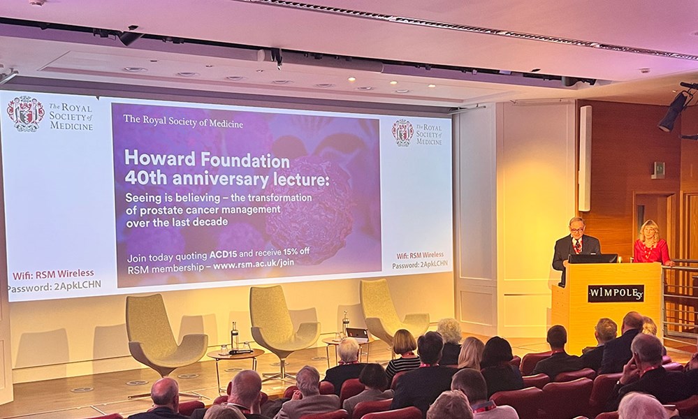
Ms Julie Lambert, Chair, Howard Foundation and Mr Jon Howard, Secretary, Howard Foundation introduce the lecture
You can watch the Howard Foundation 40th anniversary lecture in full on our YouTube channel.
The RSM would like to thank The Howard Foundation for their support of this hybrid event.